
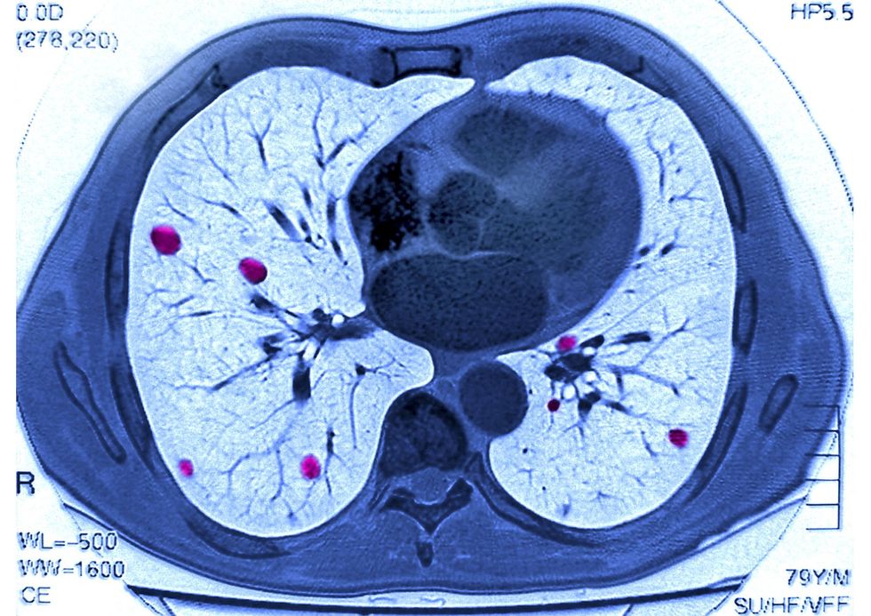
Scientists at the University of Edinburgh, working alongside NHS Lothian, have unveiled a promising new technique that could transform how doctors determine optimal treatments for lung cancer patients.
The innovative method offers a way to accurately forecast genetic alterations responsible for lung cancer development, bypassing the slower and costlier conventional laboratory approaches currently in use.
Experts believe the advancement could significantly accelerate the diagnostic process for those suffering from the disease.
By enabling clinicians to pinpoint suitable therapies more rapidly, the technology represents a potentially significant step forward for British medical science and patient care.
Lung cancer continues to claim more lives globally than any other form of the disease
|
GETTY
Lung cancer continues to claim more lives globally than any other form of the disease, according to the University of Edinburgh.
Certain lung tumours harbour specific DNA mutations that influence whether patients might respond favourably to targeted therapies.
However, identifying these genetic changes presently demands laboratory procedures such as gene sequencing.
These tests prove both expensive and lengthy, whilst also consuming precious tissue obtained from often small biopsy samples.
The constraints place considerable strain on diagnostic services, particularly as medical professionals encounter growing numbers of patients diagnosed at earlier stages of the illness.
This surge has resulted in an increasing volume of biopsy specimens requiring analysis.
The breakthrough employs fluorescence lifetime imaging microscopy, known as FLIM, which captures natural light emissions from tissue specimens before artificial intelligence analyses the resulting patterns.
During trials, the approach demonstrated remarkable precision in detecting EGFR mutations and could differentiate between the two most prevalent variants crucial for treatment planning.
“This approach has the potential to take processes that currently cost thousands of pounds and require weeks of lab work and reduce them to something that takes minutes and costs hundreds,” Dr Qiang Wang, co-lead of the study from the Institute for Regeneration and Repair, said.
“Technologies like this, which can deliver more information from smaller tissue samples at speed, will be essential for developing clinically effective diagnostic pathways,” Dr David Dorward, consultant thoracic pathologist at NHS Lothian, added.
Professor Ahsan Akram, who co-led the research from the Institute for Regeneration and Repair, expressed optimism about what lies ahead.
“This is a significant step towards a future where a single, non-destructive fluorescence scan of a biopsy could quickly inform clinicians whether a patient has cancer, what type of cancer they have and now, with this work, if it is likely to respond to targeted treatment, helping to ensure the right treatment reaches the right patient more quickly,” he said.
Scientists hope to extend the platform’s capabilities to additional cancer types and other targetable mutations,
|
GETTY
The research team is currently pursuing clinical validation of the technique.
Further efforts aim to extend the platform’s capabilities to additional cancer types and other targetable mutations, with plans to integrate the system into routine clinical practice.
